Photo from wikipedia
BackgroundDown syndrome (DS) is by far the most common known chromosomal disorder. Some characteristic features of DS are generalised growth deficiency, craniofacial abnormalities such as mandibular prognathism and underdevelopment of… Click to show full abstract
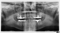
BackgroundDown syndrome (DS) is by far the most common known chromosomal disorder. Some characteristic features of DS are generalised growth deficiency, craniofacial abnormalities such as mandibular prognathism and underdevelopment of the midfacial region, dental abnormalities such as taurodontism and hypodontia. Individuals with DS have an increased prevalence of periodontal disease compared with age-matched control patients. The aim of the present study is to determine the morphologic features of the mandible among individuals with DS.MethodsThirty-four DS patients and thirty four age- and gender-matched control subjects underwent panoramic radiography, which included measurement of the mandibular canal (MC), the mandibular foramen (MF), the mandibular ramus (MR), the distance from the MC to the mandibular lower border (C-MLB), and the distance between the MC and the alveolar crest upper limit (C-AUL). Patients were separated into two groups based on age: < 15 (n = 15) and ≥ 15 (n = 19). In order to determine whether the MF, MR, MC, C-AUL, and C-MLB scores differed according to the groups (DS and control), one-way multivariate analysis of covariance (MANCOVA) was applied in which gender and age were taken as covariates.ResultsWhen the main effect according to the group was examined separately according to each measurement, the MF in the DS group was high with a moderate effect (F = 9207; p = 0.003). MR (F = 40,518; p < 0.001), MC (F = 23,747; p < 0.001), and C-AUL (F = 58,571; p < 0.001) in the DS group were lower with a larger effect. C-MLB did not significantly differ between the groups, and the effect size was quite low (p > 0.05).ConclusionsMandibular canal morphology may exhibit anatomical variations in DS. The alveolar bone level may differ from non-DS due to growth development retardation and/or periodontal diseases.
Journal Title: BMC Oral Health
Year Published: 2019
Link to full text (if available)
Share on Social Media: Sign Up to like & get
recommendations!