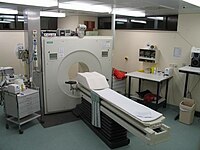
Photo from wikipedia
Background Positron emission tomography (PET) is increasingly being used as an imaging modality for clinical and research applications in veterinary medicine. Amyloid PET has become a useful tool for diagnosing… Click to show full abstract
Background Positron emission tomography (PET) is increasingly being used as an imaging modality for clinical and research applications in veterinary medicine. Amyloid PET has become a useful tool for diagnosing Alzheimer’s disease (AD) in humans, by accurately identifying amyloid-beta (Aβ) plaques. Cognitive dysfunction syndrome in dogs shows cognitive and pathophysiologic characteristics similar to AD. Therefore, we assessed the physiologic characteristics of uptake of 18F-flutemetamol, an Aβ protein-binding PET tracer in clinical development, in normal dog brains, for distinguishing an abnormal state. Static and dynamic PET images of six adult healthy dogs were acquired after 18F-flutemetamol was administered intravenously at approximately 3.083 MBq/kg. For static images, PET data were acquired at 30, 60, and 90 min after injection. One week later, dynamic images were acquired for 120 min, from the time of tracer injection. PET data were reconstructed using an iterative technique, and corrections for attenuation and scatter were applied. Regions of interest were manually drawn over the frontal, parietal, temporal, occipital, anterior cingulate, posterior cingulate, and cerebellar cortices, cerebral white matter, midbrain, pons, and medulla oblongata. After calculating standardized uptake values with an established formula, standardized uptake value ratios (SUVRs) were obtained, using the cerebellar cortex as a reference region. Results Among the six cerebral cortical regions, the cingulate cortices and frontal lobe showed the highest SUVRs. The lowest SUVR was observed in the occipital lobe. The average values of the cortical SUVRs were 1.25, 1.26, and 1.27 at 30, 60, and 90 min post-injection, respectively. Tracer uptake on dynamic scans was rapid, peaking within 4 min post-injection. After reaching this early maximum, cerebral cortical regions showed a curve with a steep descent, whereas cerebral white matter demonstrated a curve with a slow decline, resulting in a large gap between cerebral cortical regions and white matter. Conclusion This study provides normal baseline data of 18F-flutemetamol PET that can facilitate an objective diagnosis of cognitive dysfunction syndrome in dogs in future.
Journal Title: BMC Veterinary Research
Year Published: 2020
Link to full text (if available)
Share on Social Media: Sign Up to like & get
recommendations!