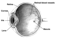
Photo from wikipedia
Extensive effort has been made studying retinal pathology in Alzheimer’s disease (AD) to improve early noninvasive diagnosis and treatment. Particularly relevant are vascular changes, which appear prominent in early brain… Click to show full abstract
Extensive effort has been made studying retinal pathology in Alzheimer’s disease (AD) to improve early noninvasive diagnosis and treatment. Particularly relevant are vascular changes, which appear prominent in early brain pathogenesis and could predict cognitive decline. Recently, we identified platelet-derived growth factor receptor beta (PDGFRβ) deficiency and pericyte loss associated with vascular Aβ deposition in the neurosensory retina of mild cognitively impaired (MCI) and AD patients. However, the pathological mechanisms of retinal vascular changes and their possible relationships with vascular amyloidosis, pericyte loss, and blood-retinal barrier (BRB) integrity remain unknown. Here, we evaluated the retinas of transgenic APP SWE /PS1 ΔE9 mouse models of AD (ADtg mice) and wild-type mice at different ages for capillary degeneration, PDGFRβ expression, vascular amyloidosis, permeability and inner BRB tight-junction molecules. Using a retinal vascular isolation technique followed by periodic acid-Schiff or immunofluorescent staining, we discovered significant retinal capillary degeneration in ADtg mice compared to age- and sex-matched wild-type mice ( P < 0.0001). This small vessel degeneration reached significance in 8-month-old mice ( P = 0.0035), with males more susceptible than females. Degeneration of retinal capillaries also progressively increased with age in healthy mice ( P = 0.0145); however, the phenomenon was significantly worse during AD-like progression ( P = 0.0001). A substantial vascular PDGFRβ deficiency (~ 50% reduction, P = 0.0017) along with prominent vascular Aβ deposition was further detected in the retina of ADtg mice, which inversely correlated with the extent of degenerated capillaries (Pearson’s r = − 0.8, P = 0.0016). Importantly, tight-junction alterations such as claudin-1 downregulation and increased BRB permeability, demonstrated in vivo by retinal fluorescein imaging and ex vivo following injection of FITC-dextran (2000 kD) and Texas Red-dextran (3 kD), were found in ADtg mice. Overall, the identification of age- and Alzheimer’s-dependent retinal capillary degeneration and compromised BRB integrity starting at early disease stages in ADtg mice could contribute to the development of novel targets for AD diagnosis and therapy.
Journal Title: Acta Neuropathologica Communications
Year Published: 2020
Link to full text (if available)
Share on Social Media: Sign Up to like & get
recommendations!