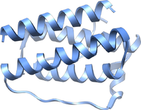
Photo from wikipedia
Adaptive thermogenesis in brown adipose tissue is stimulated by the sympathetic nervous system (SNS) in response to cold stress. Using retrograde viral transneuronal tract tracers, previous studies have identified that… Click to show full abstract
Adaptive thermogenesis in brown adipose tissue is stimulated by the sympathetic nervous system (SNS) in response to cold stress. Using retrograde viral transneuronal tract tracers, previous studies have identified that the paraventricular nucleus (PVN), ventromedial hypothalamus (VMH), and median preoptic nucleus (MnPO) contain neurons that are part of sympathetic outflow tracts to brown adipose tissue, presumptively involved in SNS stimulation of interscapular brown adipose tissue (iBAT). Pituitary Adenylate Cyclase-Activating Polypeptide (PACAP) is a peptide hormone known to regulate energy homeostasis, acting in both the central (CNS) and peripheral nervous system (PNS). Mice lacking PACAP have impaired adrenergic-induced thermogenesis and a cold-sensitive phenotype. In the CNS, PACAP is highly expressed in the VMH, MnPO, and PVN of the hypothalamus. Injection of PACAP into the VMN increased core body temperature and sympathetic nerve activity to brown adipose tissue. While these studies show exogenous PACAP can activate sympathetic outflow tracts to brown adipose tissue, they do not confirm that endogenously expressed PACAP induces sympathetic nerve activity as an adaptive mechanism to cold stress, or if sympathetic outflow tracts originating in the hypothalamus express PACAP. We hypothesize that PACAP is expressed in neurons of sympathetic outflow tracts originating in the hypothalamus. To test this hypothesis, PACAP-eGFP transgenic mice were injected with the retrograde neural tracer, pseudorabies virus tagged with β-galactosidase (β-gal, PRV-BaBlu), in iBAT where postganglionic nerves innervate the tissue. Five-days post-infection, animals were culled, brains removed and cryosectioned. Neurons positive for green fluorescent protein (eGFP) and/or β-gal immunoreactivity (ir) were identified by immunohistochemistry in serial coronal and sagittal brain cryo-sections. Co-occurrence of eGFP-ir and β-gal-ir, inferred PACAP expressing neurons present in sympathetic outflow tracts (ImageJ). Co-occurrence was identified in several structures in the hypothalamus and thalamus. In conclusion, this study presents neuroanatomical evidence for populations of PACAPinergic neurons in the hypothalamus that are part of sympathetic outflow tracts to brown adipose tissue, providing further evidence of a central role for PACAP in regulating energy homeostasis.
Journal Title: Journal of the Endocrine Society
Year Published: 2021
Link to full text (if available)
Share on Social Media: Sign Up to like & get
recommendations!