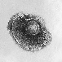
Photo from wikipedia
Objective Herein, we report three patients presenting with zoster ophthalmicus caused by varicella zoster virus (VZV) with unexpected and novel cervico-medullary findings on magnetic resonance imaging (MRI). Background Zoster opthalmicus… Click to show full abstract
Objective Herein, we report three patients presenting with zoster ophthalmicus caused by varicella zoster virus (VZV) with unexpected and novel cervico-medullary findings on magnetic resonance imaging (MRI). Background Zoster opthalmicus involves reactivation of latent VZV in the ophthalmic division of the trigeminal nerve producing a dermatomal rash. In a minority of patients this leads to ophthalmic findings and damage to surrounding peripheral nerves through perineural/intraneural inflammation. MRI findings in zoster opthalmicus are often nondescript or normal leading to misdiagnosis and delays in treatment. Design/Methods This was a case-series describing patients in Calgary, Alberta who presented with zoster opthalmicus, and who were noted incidentally to have similar appearing lesions in the ipsilateral cervico-medullary region on brain MRI. Standard MRI imaging of the brain and selected high-resolution images of the orbits and globes were reviewed by neuroradiology (CW). Results Three patients, with varying phenotypes of zoster opthalmicus, are described: a 70-yo male with ipsilateral decreased visual acuity and oculomotor/abducens palsy; a 75-yo female with ipsilateral oculomotor/facial nerve palsies; and a 35-yo immunocompromised female with ipsilateral blepharoconjunctivitis and optic neuritis. In all 3 cases, MRI revealed profound T2 and fluid-attenuated inversion recovery (FLAIR) hyperintensity along the spinotrigeminal tract of the clinically affected side, extending from the posterolateral pons and medulla into the cervical region. Conclusions To our knowledge, hyperintensity within the medulla and spinal trigeminal tract in zoster opthalmicus is a rare imaging finding. We have coined this radiologic finding the “trigeminal tract sign” due to the anatomic structure affected. A thorough radiologic/laboratory workup should be considered in patients with similar presentations; however, a high index of suspicion for zoster opthalmicus should be considered in cases with the aforementioned imaging findings. Whether this radiological finding predicts disease severity or the likelihood of multiple cranial nerve involvement (as was seen in all patients in this series) requires further study.
Journal Title: Neurology
Year Published: 2022
Link to full text (if available)
Share on Social Media: Sign Up to like & get
recommendations!