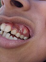
Photo from wikipedia
Background: Condylar Hyperplasia (CH) is a self-limiting mandibular condyle disorder that shows asymmetry progress conjunction with associated occlusal changes as long as condylar growth is still active and leads to… Click to show full abstract
Background: Condylar Hyperplasia (CH) is a self-limiting mandibular condyle disorder that shows asymmetry progress conjunction with associated occlusal changes as long as condylar growth is still active and leads to facial asymmetry. This study aimed to evaluate dental arches by analyzing dental arch asymmetry and form in orthodontic patients with CH in a North Sumatra subpopulation. Methods: This is a retrospective study of suspected CH patient's clinical records who sought for the initial orthodontic treatment between January 2015 to March 2019. Patient with facial asymmetry (based on photography, posterior cross bite and midline deviation), positive temporomandibular joint disorder in functional analysis, and no history of facial trauma were included in the study. Dental arch asymmetry was based on the measurement of dental midline deviation, canine tip in the dental arch, distance of the upper canines from the palatal suture, and inter canine distance. The evaluation of dental arch was achieved by comparing arch width and length. Results: There was a significant difference (p<0.05) of upper canine distance from the palatal suture in female patients when evaluating upper dental arch asymmetry. There was a moderate correlation (r=0.379) in midline deviation between upper and lower dental arch. The dimension and dental arch form was mid and flat, and there was moderate correlation (r=0.448) between the upper and lower dental arch form in these CH patients. Conclusion: The evaluation of dental arch symmetry and arch form showed asymmetric occlusal characteristics in orthodontics patient with CH in North Sumatera subpopulation. In treating these patients, we recommend the plaster cast evaluation as essential and routine procedure in order to understand the complexity of occlusal change due to active growth of condylar and limitation in radiography evaluation.
Journal Title: F1000Research
Year Published: 2020
Link to full text (if available)
Share on Social Media: Sign Up to like & get
recommendations!