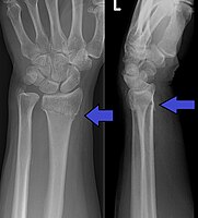
Photo from wikipedia
Purpose Current clinical and radiological methods of predicting a patient’s growth potential are limited in terms of practicality, accuracy and known to differ in different races. This information influences optimal… Click to show full abstract
Purpose Current clinical and radiological methods of predicting a patient’s growth potential are limited in terms of practicality, accuracy and known to differ in different races. This information influences optimal timing of bracing and surgical intervention in adolescent idiopathic scoliosis (AIS). The Luk classification was developed to mitigate limitations of existing tools. Few reliability studies are available and are limited to certain geographical regions with varying results. This study was performed to analyze reproducibility and reliability of the Luk Distal Radius and Ulna Classification in European patients. Methods This is a radiological study of 50 randomly selected left hand and wrist radiographs of patients with AIS referred to a tertiary referral centre. They were assessed for bone maturity using the Luk Distal Radius and Ulna Classification. Assessment was performed twice by four examiners at an interval of one month. Statistical analysis was performed using the intraclass correlation (ICC) method to determine the reliabilities within and between the examiners. Results In total, 50 radiographs (M:F = 13:37) with a mean age of 13.7 years (10 to 18) were assessed for reliability. The inter-rater ICC value was 0.918 for radius assessment and 0.939 for ulna assessment. The intra-rater ICC values for radius assessment ranged between 0.897 and 0.769 and between 0.948 and 0.786 for ulna assessment. There was near perfect correlation for both assessments. Conclusion This study provides independent evidence that the Luk Distal Radius and Ulna Classification is a reliable tool for assessment of skeletal maturity for European patients. Minimal clinical experience is required to reliably utilize it. Level of evidence IV
Journal Title: Journal of Children's Orthopaedics
Year Published: 2021
Link to full text (if available)
Share on Social Media: Sign Up to like & get
recommendations!