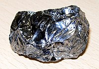
Photo from wikipedia
We present Raman analysis of nanosecond laser textured silicon. The samples have also been characterized by field emission scanning electron microscopy (FESEM) and x ray diffraction. Contact angles (CAs) are… Click to show full abstract
We present Raman analysis of nanosecond laser textured silicon. The samples have also been characterized by field emission scanning electron microscopy (FESEM) and x ray diffraction. Contact angles (CAs) are measured to trace the hydrophilic nature. Characterization of the textured samples in argon and air shows that cleavage cracks are developed during texturing. CA measurements reveal the superhydrophilic nature of textured samples obtained in the presence of ambient oxygen and argon. In vacuum, however, the hydrophilicity is decreased. Micro-Raman analysis indicates the formation of nano-sized cleavage cracks that impart stable superhydrophilic properties to textured silicon is supported from FESEM images also. On the other hand, in vacuum textured silicon, evidence of such cracks is not noticed, which is also supported by Raman analysis. Further, the hydrophilicity is decreased. A definitive trend appears to exist between Raman signatures and hydrophilicity. We believe that the study will further the understanding of the mechanistic aspect in designing textured silicon with a high degree of self-cleaning capability.
Journal Title: Applied optics
Year Published: 2022
Link to full text (if available)
Share on Social Media: Sign Up to like & get
recommendations!