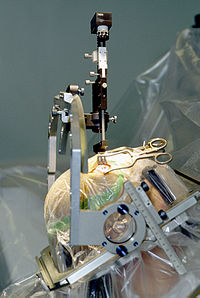
Photo from wikipedia
The rationale for this project is to evaluate the efficiency of a novel sonographic method for measurements of interosseous distances. The method utilizes a propagating ultrasonic beam through aqueous milieu… Click to show full abstract
The rationale for this project is to evaluate the efficiency of a novel sonographic method for measurements of interosseous distances. The method utilizes a propagating ultrasonic beam through aqueous milieu which is directed as a jet into a drilled tract. We used a plastic model of human L5 vertebra and ex vivo specimen of L5 porcine vertebra and generated 2 mm in diameter tracts in vertebral pedicles. The tracts were created in the “desired” central direction and in the “wrong” medial and lateral directions. The drilled tracts and the residual, up to opposite cortex, distances were measured sonographically and mechanically and compared statistically. We show that "true” mechanical measurements can be predicted from sonographic measurements with correction of 1–3 mm. The correct central route can be distinguished from the wrong misplaced routes. By using the sonographic measurements, a correct direction of drilling in the pedicle of lumbar L5 vertebra can be efficiently monitored.
Journal Title: PLoS ONE
Year Published: 2017
Link to full text (if available)
Share on Social Media: Sign Up to like & get
recommendations!