Photo from wikipedia
Background Visceral adiposity is a risk factor for many chronic diseases. Existing methods to quantify visceral adipose tissue volume using computed tomographic (CT) images often use a single slice, are… Click to show full abstract
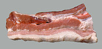
Background Visceral adiposity is a risk factor for many chronic diseases. Existing methods to quantify visceral adipose tissue volume using computed tomographic (CT) images often use a single slice, are manual, and are time consuming, making them impractical for large population studies. We developed and validated a method to accurately, rapidly, and robustly measure visceral adipose tissue volume using CT images. Methods In-house software, Medical Executable for the Efficient and Robust Quantification of Adipose Tissue (MEERQAT), was developed to calculate visceral adipose tissue volume using a series of CT images within a manually identified region of interest. To distinguish visceral and subcutaneous adipose tissue, ellipses are drawn through the rectus abdominis and transverse abdominis using manual and automatic processes. Visceral and subcutaneous adipose tissue volumes are calculated by counting the numbers of voxels corresponding to adipose tissue in the region of interest. MEERQAT’s ellipse interpolation method was validated by comparing visceral adipose volume from 10 patients’ CT scans with corresponding results from manually delineated scans. Accuracy of visceral adipose quantification was tested using a phantom consisting of animal fat and tissues. Robustness of the method was tested by determining intra-observer and inter-observer coefficients of variation (CV). Results The mean difference in visceral adipose tissue volume between manual and elliptical delineation methods was -0.54 ± 4.81%. In the phantom, our measurement differed from the known adipose volume by ≤ 7.5% for all scanning parameters. Mean inter-observer CV for visceral adipose tissue volume was 0.085, and mean intra-observer CV for visceral adipose tissue volume was 0.059. Conclusions We have developed and validated a robust method of accurately and quickly determining visceral adipose tissue volume in any defined region of interest using CT imaging.
Journal Title: PLoS ONE
Year Published: 2017
Link to full text (if available)
Share on Social Media: Sign Up to like & get
recommendations!