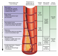
Photo from wikipedia
Activation of the classical complement pathway plays a major role in regulating atherosclerosis progression, and it is believed to have both proatherogenic and atheroprotective effects. This study focused on C1q,… Click to show full abstract
Activation of the classical complement pathway plays a major role in regulating atherosclerosis progression, and it is believed to have both proatherogenic and atheroprotective effects. This study focused on C1q, the first protein in the classical pathway, and examined its potentialities of plaque progression and instability and its relationship with clinical outcomes. To assess the localization and quantity of C1q expression in various stages of atherosclerosis, immunohistochemistry, western blotting, and real-time polymerase chain reaction (PCR) were performed using abdominal aortas from eight autopsy cases. C1q immunoreactivity in relation to plaque instability and clinical outcomes was also examined using directional coronary atherectomy (DCA) samples from 19 patients with acute coronary syndromes (ACS) and 18 patients with stable angina pectoris (SAP) and coronary aspirated specimens from 38 patients with acute myocardial infarction. C1q immunoreactivity was localized in the extracellular matrix, necrotic cores, macrophages and smooth muscle cells in atherosclerotic lesions. Western blotting and real-time PCR illustrated that C1q protein and mRNA expression was significantly higher in advanced lesions than in early lesions. Immunohistochemical analysis using DCA specimens revealed that C1q expression was significantly higher in ACS plaques than in SAP plaques. Finally, immunohistochemical analysis using thrombus aspiration specimens demonstrated that histopathological C1q in aspirated coronary materials could be an indicator of poor medical condition. Our results indicated that C1q is significantly involved in atherosclerosis progression and plaque instability, and it could be considered as one of the indicators of cardiovascular outcomes.
Journal Title: PLoS ONE
Year Published: 2022
Link to full text (if available)
Share on Social Media: Sign Up to like & get
recommendations!