Photo from wikipedia
Superior vena cava (SVC) aneurysms are rare mediastinal vascular lesions. We report a case of a 42-year-old female, who presented to the outpatient department with features suggestive of lower respiratory… Click to show full abstract
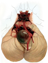
Superior vena cava (SVC) aneurysms are rare mediastinal vascular lesions. We report a case of a 42-year-old female, who presented to the outpatient department with features suggestive of lower respiratory tract infection. Chest X-ray showed abnormal contour and widening of right border of mediastinum. Computed tomography (CT) thorax revealed fusiform aneurysmal ectasia of SVC measuring 4.5 × 5.5 × 8.9 cm without internal thrombosis or dissecting flap. Management options include observation with follow-up and in some cases anticoagulation and surgical excision may be considered. The general consensus is that fusiform variety can be managed conservatively in view of the low risk of complications. The saccular aneurysms may need to be managed with anticoagulation therapy or surgically in view of the possible risk for thrombus formation and pulmonary embolism. Since in our case it was an asymptomatic primary fusiform SVC aneurysm, patient was advised for conservative management and follow-up.
Journal Title: Cardiology Research
Year Published: 2017
Link to full text (if available)
Share on Social Media: Sign Up to like & get
recommendations!