Photo from wikipedia
J-wave syndromes refer to a group of conditions of inherited arrhythmia disorders that can present with sudden cardiac death due to polymorphic ventricular arrhythmias (1). In some cases, the only… Click to show full abstract
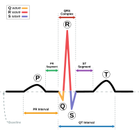
J-wave syndromes refer to a group of conditions of inherited arrhythmia disorders that can present with sudden cardiac death due to polymorphic ventricular arrhythmias (1). In some cases, the only clue to the diagnosis of these infrequent conditions is surface electrocardiogram (ECG) (2). One important point that all physicians should keep in mind is that only the presence of the ECG pattern does not constitute the syndrome (2). Several individuals will develop the ECG pattern (either spontaneously or upon certain clinical conditions), and their risk of sudden death may not differ from their counterparts without the ECG manifestation. The most commonly investigated and extensively reported J-wave syndromes in the literature are Brugada syndrome (BrS) (3) and early repolarization syndrome (ErS) (2). Both can affect young individuals with no prior history of cardiovascular disease, can coexist with “apparently” normal hearts (on initial cardiac screening including echocardiogram), and are characterized by J-point elevation (1-3). The morphology of the ST-segment and T-wave following the J-point elevation helps physicians to electrocardiographically differentiate one from the other (4). A consensus on BrS (3) has classified the type of ECG phenotypes into two variants (type-1 or “coved” and type-2 or “saddleback”), and a more recent consensus on ErS (2) has characterized the early repolarization pattern into “malignant” (more frequently associated with ErS) and “benign” (frequently seen in healthy individuals and athletes), which could be considered as a variant of normality. In the last volume of the Anatolian Journal of Cardiology, Altunbaş et al. (5) reported a remarkable case of a 68-year-old female presenting for knee surgery. She had no history of cardiovascular disease, and two recent angiograms showed no coronary artery disease. The surface ECG depicted massive J-point elevation in leads V1-V6, II, and aVF with a classic type2 Brugada ECG pattern in leads V1-V2. The combination of both patterns in the same individual was anecdotally identified as a possible marker of higher risk by McIntyre et al. (6). The case presented by Altunbaş et al. (5) resembles cases previously identified at a higher risk of developing dramatic arrhythmic events, thus indicating the need to use all diagnostic alternatives to determine the predisposition to suffer from these fatal events. As the second presented ECG normalized the pattern in leads V1-V2 (intermittency is a characteristic of BrS), a possible approach would be to challenge the sodium channels with a classic sodium channel blocker (flecainide, procainamide, or ajmaline). A positive result (induction of type-1 Brugada ECG pattern) would be confirmatory of a sodium channel dysfunction, thus helping in the management of the case. A complete genetic panel would be also desirable. A positive result may help in the screening of relatives, whereas a negative result would not rule out the condition. The follow-up of this case should include long-term cardiac monitoring; in order to detect possible ECG changes and/or self-limited episodes of polymorphic arrhythmias. If detected, appropriate therapy may help preventing possible fatal events. The use of an implantable loop recorder should be considered (7). Altunbaş et al. (5) need to be commended for the report of this impressive case; I hope to see a future report about the follow-up of this exceptional case in the next few years.
Journal Title: Anatolian Journal of Cardiology
Year Published: 2018
Link to full text (if available)
Share on Social Media: Sign Up to like & get
recommendations!