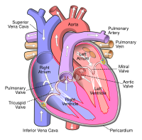
Photo from wikipedia
A 3-year-old girl with a diagnosis of tetralogy of Fallot on transthoracic echocar-diography underwent computed tomography (CT) angiography as part of pre-surgical evaluation. Incidentally, a duplicated left brachiocephalic vein (LBCV)… Click to show full abstract
A 3-year-old girl with a diagnosis of tetralogy of Fallot on transthoracic echocar-diography underwent computed tomography (CT) angiography as part of pre-surgical evaluation. Incidentally, a duplicated left brachiocephalic vein (LBCV) was observed where an accessory branch of the LBCV was seen coursing posteroinferior to the aorta in addition to a normally coursing LBCV. The accessory branch of the LBCV was thinner in caliber and seen joining the superior caval vein more caudally, at the level of azygous venous drainage. The 2 branches of the LBCV were seen encircling the aorta to join the superior caval vein at different levels resulting in a “circumaortic duplicated LBCV” (Figure 1). The aortic arch was right-sided with mirror image branching of the arch vessels. In the setting of the usual viscero-atrial arrangement, the LBCV crosses supero-anterior to the aortic arch and supra-aortic branches while coursing obliquely downward toward its confluence with the right brachiocephalic vein to form the superior caval vein. 1 Uncommonly, it can have an aberrant subaortic (or retroaor-tic) course, coursing beneath the aortic arch to drain into the superior caval vein at or caudal to the level of azygous venous drainage. 2 A duplicated LBCV encircling the aorta, where an accessory branch of LBCV having a subaortic course is present along with a normally coursing LBCV, is extremely rare and is postulated to develop secondary to persistence of both ventral and dorsal transverse anas-tomosis between the bilateral precardinal veins. 3-6 While the anomalous anatomy
Journal Title: Anatolian journal of cardiology
Year Published: 2023
Link to full text (if available)
Share on Social Media: Sign Up to like & get
recommendations!