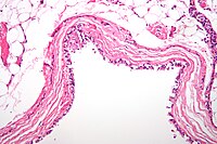
Photo from wikipedia
FIGURE 1: Coronal abdominal computed tomography (CT) scan (A) reveals a giant peritoneal hydatid cyst (arrowheads) and a calcified hepatic cyst (yellow arrow). Axial pelvic CT scans (B and C)… Click to show full abstract
FIGURE 1: Coronal abdominal computed tomography (CT) scan (A) reveals a giant peritoneal hydatid cyst (arrowheads) and a calcified hepatic cyst (yellow arrow). Axial pelvic CT scans (B and C) reveal multiple dilated and tortuous pelvic venous structures (circle) in the presacral area. A 53-year-old woman without a history of chronic disease was admitted to our hospital. On admission, the patient recounted a history of progressive abdominal distension and pelvic pain over the preceding 18 months. She had no history of systemic disease or abdominal trauma. On physical examination, a large, round abdominal mass was palpable. Abdominopelvic computed tomography revealed a giant peritoneal hydatid cyst and tortuous pelvic venous structures associated with compression by the peritoneal cyst (Figure 1).
Journal Title: Revista da Sociedade Brasileira de Medicina Tropical
Year Published: 2022
Link to full text (if available)
Share on Social Media: Sign Up to like & get
recommendations!