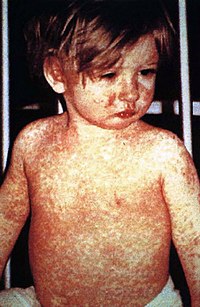
Photo from wikipedia
FIGURE 1: Ulcerated lesion with necrosis and bone exposure. A 73-year-old male rural worker from the Brazilian Amazon region presented with a 4-year left facial ulceration, which became painful as… Click to show full abstract
FIGURE 1: Ulcerated lesion with necrosis and bone exposure. A 73-year-old male rural worker from the Brazilian Amazon region presented with a 4-year left facial ulceration, which became painful as it increased in size. Physical examination revealed a deep 8-cm ulcer in the left malar and periorbital regions, associated with necrosis and purulent exudation (Figure 1). Skull computed tomography showed an osteolytic and infiltrative lesion in the malar and frontal regions (Figure 2).
Journal Title: Revista da Sociedade Brasileira de Medicina Tropical
Year Published: 2021
Link to full text (if available)
Share on Social Media: Sign Up to like & get
recommendations!