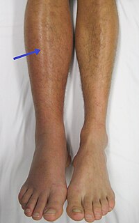
Photo from wikipedia
Cerebral venous thrombosis (CVT) is an uncommon condition that is potentially reversible if properly diagnosed and promptly treated. Although CVT can occur at any age, it most commonly affects neonates… Click to show full abstract
Cerebral venous thrombosis (CVT) is an uncommon condition that is potentially reversible if properly diagnosed and promptly treated. Although CVT can occur at any age, it most commonly affects neonates and young adults. Clinical diagnosis is difficult because the clinical manifestations of CVT are nonspecific, including headache, seizures, decreased level of consciousness, and focal neurologic deficits. Therefore, imaging is crucial for the diagnosis. Radiologists should be able to identify the findings of CVT and to recognize potential imaging pitfalls that may lead to misdiagnosis. Thus, the appropriate treatment (anticoagulation therapy) can be started early, thereby avoiding complications and unfavorable outcomes.
Journal Title: Radiologia Brasileira
Year Published: 2022
Link to full text (if available)
Share on Social Media: Sign Up to like & get
recommendations!