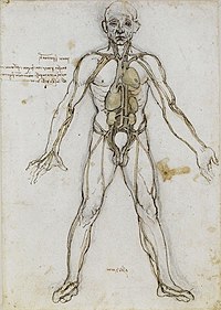
Photo from wikipedia
The mammalian ureter consists of a mesenchymal wall composed of smooth muscle cells and surrounding fibrocytes of the tunica adventitia and the lamina propria and an inner epithelial lining composed… Click to show full abstract
The mammalian ureter consists of a mesenchymal wall composed of smooth muscle cells and surrounding fibrocytes of the tunica adventitia and the lamina propria and an inner epithelial lining composed of layers of basal, intermediate, and superficial cells. How these cell types arise from multipotent progenitors is poorly understood. Here, we performed marker analysis, cell proliferation assays, and genetic lineage tracing to define the lineage relations and restrictions of the mesenchymal and epithelial cell types in the developing and mature mouse ureter. At embryonic day (E) 12.5, the mesenchymal precursor pool began to subdivide into an inner and outer compartment that began to express markers of smooth muscle precursors and adventitial fibrocytes, respectively, by E13.5. Smooth muscle precursors further diversified into lamina propria cells directly adjacent to the ureteric epithelium and differentiated smooth muscle cells from E16.5 onwards. Uncommitted epithelial progenitors of the ureter differentiated into intermediate cells at E14.5. After stratification into two layers at E15.5 and three cell layers at E18.5, intermediate cells differentiated into basal cells and superficial cells. In homeostasis, proliferation of all epithelial and mesenchymal cell types remained low but intermediate cells still gave rise to basal cells, whereas basal cells divided only into basal cells. These studies provide a framework to further determine the molecular mechanisms of cell differentiation in the tissues of the developing ureter.
Journal Title: Journal of the American Society of Nephrology : JASN
Year Published: 2017
Link to full text (if available)
Share on Social Media: Sign Up to like & get
recommendations!