Photo from wikipedia
Diagnosis of non-facial melanomas on sun-damaged skin or extrafacial lentigo maligna is challenging. To identify the evolutionary dermoscopic signs, characteristic of this type of non-facial melanoma on sun-damaged skin. This… Click to show full abstract
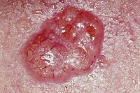
Diagnosis of non-facial melanomas on sun-damaged skin or extrafacial lentigo maligna is challenging. To identify the evolutionary dermoscopic signs, characteristic of this type of non-facial melanoma on sun-damaged skin. This retrospective descriptive observational study included 90 dermoscopic follow-up images of 22 non-facial melanomas on sun-damaged skin from 17 high-risk melanoma patients, followed with digital dermoscopy and diagnosed between January 2016 and October 2020. We recorded dermoscopic changes by comparing each dermoscopic image with the previous one (mean dermoscopic follow-up of the excised lesions was 3.6 years). Confocal microscopy images were taken at diagnosis. In total, 51.5% (95% CI: 39–64) showed an appearance or increase in featureless areas with surrounding small round or triangular dark brown-blue structures, 23% (95% CI: 23-46) showed an increase in other geometric structures (angulated lines, zig-zag lines and polycyclic structures), 5.9% (95% CI: 2-14) showed an appearance or increase in bright white lines and atypical vascularization, 26.5% (95% CI: 17-39) showed an appearance or increase in follicular pigmentation structures or follicular radial lines, and 39.7% (95% CI: 28-52) showed focal islands of pigmentation in these areas. Of the changes, 54% occurred at the last and diagnostic visit. There was an increase in size in only 20.6% (95% CI: 12-32). Also, 81.8% showed pagetoid cells in the epidermis, 95.5% atypical cells at the dermoepidermal junction by reflectance confocal microscopy, and 95.5% showed non-edged or edged and non-edged papillae. This study identifies the dermoscopic evolutionary changes associated with extrafacial lentigo maligna.
Journal Title: European Journal of Dermatology
Year Published: 2022
Link to full text (if available)
Share on Social Media: Sign Up to like & get
recommendations!