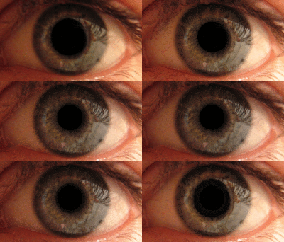
Photo from wikipedia
This review presents basic information about the state of corneal nerve fibers and Langerhans cells before and after keratoplasty. Keratoplasty is a common corneal surgery that carries a risk of… Click to show full abstract
This review presents basic information about the state of corneal nerve fibers and Langerhans cells before and after keratoplasty. Keratoplasty is a common corneal surgery that carries a risk of graft rejection. The state of corneal nerve fibers can vary after different types of keratoplasty. Corneal confocal microscopy allows in vivo evaluation of the cornea, which can help assess the condition of corneal nerve fibers, as well as reveal the presence of Langerhans cells. Further research in this direction would contribute to identifying the relationship between the state of corneal nerve fibers, the presence of Langerhans cells, and graft rejection.
Journal Title: Vestnik oftalmologii
Year Published: 2022
Link to full text (if available)
Share on Social Media: Sign Up to like & get
recommendations!