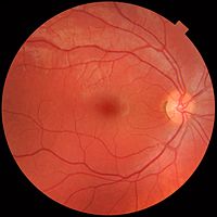
Photo from wikipedia
Diagnosing pathologies of the ocular fundus and performing differential diagnosis of intraocular tumors along with conventional ophthalmoscopy can involve additional visualization methods such as ultrasonography, fluorescein angiography and optical coherence… Click to show full abstract
Diagnosing pathologies of the ocular fundus and performing differential diagnosis of intraocular tumors along with conventional ophthalmoscopy can involve additional visualization methods such as ultrasonography, fluorescein angiography and optical coherence tomography (OCT). Many researchers note the importance of employing a multimodal approach in differential diagnosis of intraocular tumors, but there is no universally established algorithm for a rational choice of both the combination of visualizing methods, and the sequence of their application with consideration of the ophthalmoscopy findings and the results of first-line diagnostic methods. The article presents author's own multimodal algorithm developed for differential diagnosis of tumors and tumor-like diseases of the ocular fundus. This approach involves the use of such methods as OCT and Multicolor fluorescence imaging, with exact sequence and combination determined on the basis of ophthalmoscopy and ultrasonography findings.
Journal Title: Vestnik oftalmologii
Year Published: 2023
Link to full text (if available)
Share on Social Media: Sign Up to like & get
recommendations!