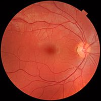
Photo from wikipedia
AIM To compare the assessment outcomes of the characteristics of mild to moderate non-proliferative diabetic retinopathy (NPDR) established by fundus photography and fundus fluorescein angiography (FFA). METHODS The fundus photos… Click to show full abstract
AIM To compare the assessment outcomes of the characteristics of mild to moderate non-proliferative diabetic retinopathy (NPDR) established by fundus photography and fundus fluorescein angiography (FFA). METHODS The fundus photos and FFA results of 260 patients with diabetes mellitus were reviewed. Diabetic retinopathy (DR) severity was graded based on the international classification standard. The microaneurysms, hemorrhages, and intraretinal microvascular abnormalities (IRMA) in FFA images of patients with mild to moderate NPDR were observed. The differences between the fundus photos and the FFA results were summarized, analyzed, and compared. RESULTS The counting of intraretinal hemorrhages identified by FFA revealed that only 9 eyes (1.9%) had more than 20 intraretinal hemorrhages in all four quadrants; 15 eyes (3.1%) had more than 20 intraretinal hemorrhages in three quadrants; 26 eyes (5.4%) had over 20 intraretinal hemorrhages in two quadrants; and 37 eyes (7.7%) had more than 20 intraretinal hemorrhages in only one quadrant. Furthermore, the number of IRMAs appeared ≥4 in 17 eyes, 3 in 35 eyes, 2 in 69 eyes, and 1 in 93 eyes. CONCLUSION FFA has higher detection accuracy of retinal angiopathy than fundus photography. FFA grading results are helpful for timely detection and proper treatment of lesions easily missed by fundus photography.
Journal Title: International journal of ophthalmology
Year Published: 2022
Link to full text (if available)
Share on Social Media: Sign Up to like & get
recommendations!