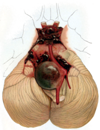
Photo from wikipedia
Unruptured brain arteriovenous malformations (AVM) have a heterogeneous clinical presentation, mainly related to the presence of intracerebral hemorrhage. We report the diagnosis of AVM in a patient with Parkinson's disease… Click to show full abstract
Unruptured brain arteriovenous malformations (AVM) have a heterogeneous clinical presentation, mainly related to the presence of intracerebral hemorrhage. We report the diagnosis of AVM in a patient with Parkinson's disease (PD), who undergone positron emission tomography/magnetic resonance imaging (PET/MRI) molecular brain imaging with fluorine-18-dihydroxyphenylalanine (18F-DOPA). This case suggests that AVM may be occasionally recognized in molecular imaging studies using positron-emitting amino acids. Magnetic resonance imaging with susceptibility-weighted imaging (SWI) sequences and 3D time of flight (TOF) reconstruction may contribute to manage patients with AVM.
Journal Title: Hellenic journal of nuclear medicine
Year Published: 2022
Link to full text (if available)
Share on Social Media: Sign Up to like & get
recommendations!