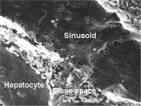
Photo from wikipedia
Fibrosis, defined as the excessive accumulation of extracellular matrix proteins, is a key feature in most chronic inflammatory diseases (1). Fibrosis can affect nearly all tissues and organs in the… Click to show full abstract
Fibrosis, defined as the excessive accumulation of extracellular matrix proteins, is a key feature in most chronic inflammatory diseases (1). Fibrosis can affect nearly all tissues and organs in the body. While fibrosis is typically reversible, for example as part of normal wound healing, it can become irreversible when the tissue injury is chronic, severe or repetitive. Permanent scarring can lead to organ failure and ultimately even to death. It is therefore of key importance that patients are routinely monitored to evaluate the severity of fibrosis for effective management of their disease. The reference standard for detection and staging of fibrosis is pathological sampling. However, next to being invasive, needle biopsy procedures only sample a small part of the tissue, while fibrogenesis has shown to be a highly heterogenous process (2). Therefore, there is increasing interest in the development of noninvasive imaging methods for assessment of fibrosis. From the evaluated imaging techniques, elastography methods measuring tissue stiffness have proved particularly promising to evaluate fibrosis, especially in the liver (3). However, an increase in stiffness is not specific to fibrosis, as other pathological changes including inflammation may also increase tissue stiffness (4). In addition, elastography methods require propagation of external mechanical waves into the tissue of interest. Wave propagation may be limited in obese patients or when applying the elastography method for imaging of organs located deeper in the body. Thus, there remains a need for an alternative imaging method that is more specific to fibrosis and is more widely applicable in all patients and all organs. The paper by Zhao et al. (5) evaluates the sensitivity of MRI relaxation parameter T1ρ to liver fibrosis in a rat model of non-alcoholic fatty liver disease (NAFLD). T1ρ, defined as the longitudinal relaxation time in the rotating frame, is a measure of the decay of magnetization in the transverse plane in the presence of a spin-lock pulse that is applied parallel to the magnetization vector. As T1ρ is sensitive to lowfrequency interactions between macromolecules and bulk water, there has been significant interest in application of T1ρ for measurement of collagen deposition in fibrotic tissues, including in the liver (6,7), kidney (8), myocardium (9) and spleen (10). A significant positive correlation of collagen content with T1ρ has been observed in both kidney (8) and liver tissues (11). While T1ρ has consistently shown an increase in the presence of fibrosis (6-8,11), the exact mechanism of this T1ρ elevation has not been determined. Intuitively, one would expect a decrease in T1ρ in fibrotic tissues, due to increased interactions between extracellular matrix proteins and water protons. The observed increase in T1ρ may therefore be related to other concomitant pathological processes, including inflammation and steatosis in the context of liver fibrosis. The elegant design of the study by Zhao et al. allowed for more detailed elucidation of the pathological changes that drive T1ρ elevation in NAFLD. MRI and histopathological evaluation of the liver were performed at several time points during methionine and choline-deficient (MCD) diet in the NAFLD group. A separate control group was also included. This study design allowed for separate analysis of the influence of fibrosis, inflammation and steatosis on liver T1ρ. A highly significant positive correlation was found between collagen content and liver T1ρ (r=0.82, P<0.0001), Editorial Commentary
Journal Title: Quantitative imaging in medicine and surgery
Year Published: 2020
Link to full text (if available)
Share on Social Media: Sign Up to like & get
recommendations!