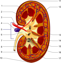
Photo from wikipedia
Background For complicated Stanford type B aortic dissection (TBAD), thoracic endovascular aortic repair (TEVAR) is the recommended treatment; however, the type of renal artery that should be repaired remains controversial.… Click to show full abstract
Background For complicated Stanford type B aortic dissection (TBAD), thoracic endovascular aortic repair (TEVAR) is the recommended treatment; however, the type of renal artery that should be repaired remains controversial. The study aimed to investigate the changes in the renal artery and renal volume in complicated TBAD after TEVAR and the predictors of renal atrophy. Methods The cohort study retrospectively enrolled patients with acute and subacute complicated TBAD who underwent aortic computed tomography angiography (CTA) 1 month before as well as 1 week and half a year after TEVAR from January 2010 to May 2017. According to the source of blood supply shown in preoperative CT, the renal artery was classified in 3 ways: type 1, supplied by the aortic true lumen; type 2, supplied by the aortic false lumen; or type 3, supplied by both the true and false lumen. Results A total of 91 patients (81 men and 10 women) with an average age of 48.12±10.35 years were enrolled. Renal arteries were classified as type 1 (n=91), type 2 (n=35), and type 3 (n=56). There was no difference in the distribution of the 3 types on the left and right sides (type 1 vs. type 2 vs. type 3: 52:39 vs. 15:20 vs. 24:32; P=0.152). After TEVAR, type 3 was more likely to have spontaneous healing than type 2 (16.1% vs. 2.9%; P=0.049). There was no significant difference in the preoperative volume of kidneys of the 3 types (type 1 vs. type 2 vs. type 3: 198.23±38.68 vs. 197.37±41.77 vs. 195.10±36.11 mL; P=0.893). The postoperative volume of types 2 and 3 was smaller than that of type 1 (type 1 vs. type 2 vs. type 3: 190.09±43.25 vs. 165.15±52.63 vs. 170.70±45.28 mL; P=0.006). The renal volume was reduced in all 3 types of renal artery, especially in type 2 (the change of renal volume for type 1 vs. type 2 vs. type 3: −8.14±29.31 vs. −32.22±41.59 vs. −24.41±38.44 mL; P=0.001). The relative change of renal volume for type 1 vs. type 2 vs. type 3: (−3.64±15.69)% vs. (−16.00±21.29)% vs. (−11.97±18.22)%; P=0.001). During the median follow-up of 668 days, 7 patients (7.7%) belonging to types 2 and 3 developed renal atrophy. False lumen thrombosis in the abdominal aorta and/or the renal artery was the predictor of renal atrophy [hazard ratio (HR) =17.757; P=0.008]. Conclusions Patients with type 2 or 3 renal artery and false lumen thrombosis in the abdominal aorta and/or renal artery should be monitored closely and actively intervened to prevent renal atrophy.
Journal Title: Quantitative Imaging in Medicine and Surgery
Year Published: 2022
Link to full text (if available)
Share on Social Media: Sign Up to like & get
recommendations!