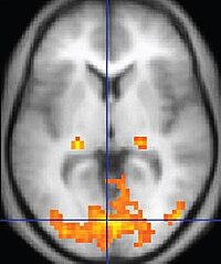
Photo from wikipedia
This paper updates and extends three previous papers on tissue property filters (TP-filters), Multiplied, Added, Divided and/or Subtracted Inversion Recovery (MASTIR) pulse sequences and synergistic contrast MRI (scMRI). It does… Click to show full abstract
This paper updates and extends three previous papers on tissue property filters (TP-filters), Multiplied, Added, Divided and/or Subtracted Inversion Recovery (MASTIR) pulse sequences and synergistic contrast MRI (scMRI). It does this by firstly adding the central contrast theorem (CCT) to TP-filters, secondly including division with MASTIR sequences to make them Multiplied, Added, Subtracted and/or Divided IR (MASDIR) sequences, and thirdly incorporating division into the image processing needed for scMR to increase synergistic T1 contrast. These updated concepts are then used to explain and improve contrast at tissue boundaries, as well as to develop imaging regimes to detect and monitor small changes to the brain over time and quantify T1. The CCT is in two parts: the first part states that contrast produced by each TP is the product of the change in TP multiplied by the TP sequence weighting which is the first partial derivative of the TP-filter. The second part states that the overall fractional contrast is the algebraic sum of the fractional contrasts produced by each of the TPs. Subtraction of two IR sequences alone about doubles contrast relative to a conventional single IR sequence. Division of this subtraction can amplify contrast 5–15 times compared with conventional IR sequences. Dividing sequences can be problematic in areas where the signal is zero but this is avoided by dividing the difference in signal of two magnitude reconstructed IR sequences by the sum of their signals. The basis for the production of high contrast, high spatial resolution boundaries at white-gray matter junctions, between cerebral cortex and cerebrospinal fluid (CSF) and at other sites with subtracted IR (SIR) and divided subtracted IR (dSIR) sequences is explained and examples are shown. A key concept is the tissue fraction f, which is the proportion of a tissue in a mixture of two tissues within a voxel. Contrast at boundaries is a function of the partial derivative of the TP-filter, the partial derivative of the relevant TP with respect to f, and the partial derivative of f with respect to distance, x. Location of tissue boundaries is important for segmentation and is helpful in determining if inversion times have been chosen correctly. In small change regimes, the high sensitivity to small changes in T1 provided by dSIR images, together with the high definition boundaries, afford mechanisms for detecting small changes due to contrast agents, disease, perfusion and other causes. 3D isotropic rigid body registration provides a technique for following these changes over time in serial studies. Images showing high lesion contrast, high definition tissue and fluid boundaries, and the detection of small changes are included. T1 maps can be created by linearly scaling dSIR images.
Journal Title: Quantitative Imaging in Medicine and Surgery
Year Published: 2022
Link to full text (if available)
Share on Social Media: Sign Up to like & get
recommendations!