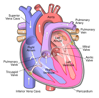
Photo from wikipedia
A 94-year-old man with long-term chronic heart failure and angina presented with a 5-day history of appetite loss and malaise. Although he had neither chest pain nor elevated cardiac enzymes,… Click to show full abstract
A 94-year-old man with long-term chronic heart failure and angina presented with a 5-day history of appetite loss and malaise. Although he had neither chest pain nor elevated cardiac enzymes, he had a high brain natriuretic peptide level of 3,450 pg/mL. An echocardiogram on admission showed moderate aortic stenosis and left ventricular severe diffuse hypokinesia with an ejection fraction of 10-20%. Therefore, we made the diagnosis of acute exacerbation of chronic heart failure. An echocardiogram on the 10th hospital day showed a 2-cm-diameter mobile left ventricular thrombus (LVT) between the posterior mitral leaflet and left ventricular lateral wall (Picture). A total of 83.3% of LVTs are formed in the apex (1). LVTs found between the lateral wall and the posterior mitral valve are extremely rare, with only one previous report existing (2). The authors state that they have no Conflict of Interest (COI).
Journal Title: Internal Medicine
Year Published: 2020
Link to full text (if available)
Share on Social Media: Sign Up to like & get
recommendations!