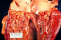
Photo from wikipedia
A 45-year old man was referred to our hospital because of a fever. His blood culture revealed a Streptococcus pneumoniae infection; ultrasound cardiography recorded vegetation at the aortic valve. Infective… Click to show full abstract
A 45-year old man was referred to our hospital because of a fever. His blood culture revealed a Streptococcus pneumoniae infection; ultrasound cardiography recorded vegetation at the aortic valve. Infective endocarditis was diagnosed, and antibiotic therapy was initiated. Although the infection and heart failure were controlled, at approximately two weeks after the antibiotic therapy initiation, a seconddegree atrioventricular block was observed. Transesophageal echocardiography revealed an annular abscess extending to the non-coronary cusp annulus (Picture 1, 2) that also communicated with the left atrium (Picture 3). We performed abscess debridement, annulus and defect reconstruction with a bovine pericardium patch, and aortic valve replacement using a mechanical valve. The patient recovered without any recurrent infective endocarditis or heart failure symptoms. Even today, annular abscesses are serious complications of infective endocarditis (1). It is important to select an appropriate treatment strategy, including surgical planning, in order to precisely diagnose the existence and extension of the abscess preoperatively.
Journal Title: Internal Medicine
Year Published: 2017
Link to full text (if available)
Share on Social Media: Sign Up to like & get
recommendations!