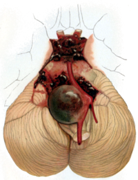
Photo from wikipedia
Recent basic studies have clarified that aneurysmal wall inflammation plays an important role in the pathophysiology of intracranial aneurysms. However, it remains an interdisciplinary challenge to visualize aneurysm wall status… Click to show full abstract
Recent basic studies have clarified that aneurysmal wall inflammation plays an important role in the pathophysiology of intracranial aneurysms. However, it remains an interdisciplinary challenge to visualize aneurysm wall status in vivo. MR-vessel wall imaging (VWI) is a current topic of advanced imaging techniques since it could provide an additional value for unruptured intracranial aneurysms (UIAs) risk stratification. With regard to ruptured intracranial aneurysms, VWI could identify a ruptured aneurysm in patients with multiple intracranial aneurysms. Intraluminal thrombus could be a clue to interpret aneurysm wall enhancement on VWI in ruptured intracranial aneurysms. The interpretation of VWI findings in UIAs would require much caution. Actually aneurysm wall enhancement in VWI was significantly associated with consensus morphologic risk factors. However, aneurysmal wall with contrast enhancement oftentimes associated with atherosclerotic, degenerated and thickened wall structure. It remains ill defined if thin wall without wall enhancement (oftentimes invisible in VWI) could be actually safe or look over wall vulnerability. We reviewed currently available studies, especially focusing on VWI for intracranial aneurysms and discussed the clinical utility of VWI.
Journal Title: Neurologia medico-chirurgica
Year Published: 2019
Link to full text (if available)
Share on Social Media: Sign Up to like & get
recommendations!