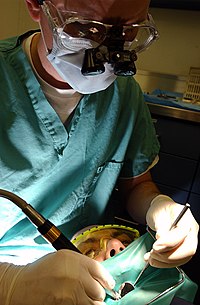
Photo from wikipedia
Dens invaginatus (DI) is a developmental anomaly that poses a significant challenge to the clinician if endodontic treatment is required. The type II (as per Oehlers) form exhibits complex internal… Click to show full abstract
Dens invaginatus (DI) is a developmental anomaly that poses a significant challenge to the clinician if endodontic treatment is required. The type II (as per Oehlers) form exhibits complex internal anatomy and is frequently associated with incomplete root and apex formation. The purpose of this study is to present two cases of type II DI in the maxillary lateral incisors. In the first case, non-surgical endodontic therapy was performed utilizing calcium hydroxide as an intracanal dressing, showing significant periapical healing of the apical radiolucent area at the six month follow-up. In the second case, the development of the root and apex were affected by pulp necrosis, and the revascularization procedure was performed. Complete resolution of the pre-existing apical radiolucency, apical closure, thickening of the root canal walls, and increase in root length, after 32 months was observed. Early detection of teeth with DI type II and proper exploration of their internal anatomy are key factors for their successful management. As demonstrated in this report, conservative non-surgical endodontic treatment should be the first line of treatment for these cases. The use of revascularization protocols in teeth that develop pulp necrosis and exhibit early stage of root development could be a better alternative than traditional apexification techniques.
Journal Title: Iranian endodontic journal
Year Published: 2017
Link to full text (if available)
Share on Social Media: Sign Up to like & get
recommendations!