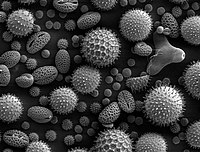
Photo from wikipedia
Pebrine disease is the most important and dangerous disease of silkworm caused by Nosema bombycis as an obligate intracellular parasitic fungus. It has caused tremendous economic losses in the silk… Click to show full abstract
Pebrine disease is the most important and dangerous disease of silkworm caused by Nosema bombycis as an obligate intracellular parasitic fungus. It has caused tremendous economic losses in the silk industry in recent years. Given the fact that light microscopy method (with low accuracy) is the only method for diagnosing pebrine disease in the country, transmission electron microscopy (TEM) and scanning electron microscopy (SEM) methods were adopted in this study for accurate morphological identification of the spores causing pebrine disease. Infected larvae and mother moth samples were collected from several farms (Parand, Parnian, Shaft, and Iran Silk Research Center in Gilan province, Iran). The spores were then purified using the sucrose gradient method. From each region, 20 and 10 samples were prepared for SEM and TEM analysis, respectively. In addition, an experiment was performed to evaluate the symptoms of pebrine disease by treating fourth instars with the spores purified for the present study, along with a control group. The results of SEM analysis showed that the mean±SD length and width of spores were 1.99±0.25 to 2.81±0.32 μm, respectively. Based on the obtained results, the size of spores was smaller than the Nosema bombycis (N. bombycis) as the classic species that cause pebrine disease. In addition, transmission electron microscopy (TEM) pictures showed that the grooves of the adult spores were deeper than those of other Nosema species, Vairomorpha, and Pleistophora, and resembled N. bombycis in other studies. Examination of pathogenicity of the studied spores indicated that the disease symptoms in controlled conditions were similar to those in the sampled farms. The most important symptom in fourth and fifth instrars were the small size and no growth in the treatment group compared with the control group. Findings of SEM and TEM analysis showed better morphological and structural details of parasite compared with light microscopy, and demonstrated that the studied species were a native strain of N. bombycis specific to Iran, whose size and other characteristics were unique and introduced for the first time in this study.
Journal Title: Archives of Razi Institute
Year Published: 2022
Link to full text (if available)
Share on Social Media: Sign Up to like & get
recommendations!