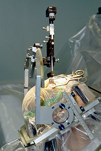
Photo from wikipedia
Indocyanine green video angiography (ICG-VA) is a non-invasive, easy to use and very useful tool for various neurosurgical procedures. The first application was in neurovascular surgery, because it was born… Click to show full abstract
Indocyanine green video angiography (ICG-VA) is a non-invasive, easy to use and very useful tool for various neurosurgical procedures. The first application was in neurovascular surgery, because it was born as an intravascular tracer for vessels visualization; this has been really useful in aneurysms, AVMs and dural fistulas surgery where identification, obliteration or potency of vessels is essential. Introduced in vascular neurosurgery since 2003, ICG-VA applications have broadened over time, both in vascular and in other neurosurgical fields. As the first, in 2003 Raabe et al. [1] described the use of ICG-VA for intraoperative assessment of cerebral vascular flow, enabling visualization of vessel potency and aneurysm occlusion during aneurysm surgery. ICG-VA applications in vascular neurosurgery have significantly increased over time including complex aneurysms, bypass, atero-venous malformations (AVM) arterovenous fistulas (AVF), evaluation of cortical perfusion.[2-4]. The procedure can be easily repeated after 5-10min. Adverse reactions are comparable to those of other types of contrast media, with frequencies of 0.05% (hypotension, arrhythmia, or, more rarely, anaphylactic shock) to 0.2% (nausea, pruritus, syncope, or skin eruptions. The aim of the present study is to systematically analyses ICG-VA applications in vascular neurosurgery, highlighting the reported advantages and disadvantages, and discussing future perspectives.
Journal Title: Journal of neurosurgical sciences
Year Published: 2019
Link to full text (if available)
Share on Social Media: Sign Up to like & get
recommendations!