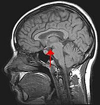
Photo from wikipedia
Abstract Objective. The aim of the present study was to find out whether acute effect of different doses of selected antipsychotics including aripiprazole (ARI), amisulpride (AMI), asenapine (ASE), haloperidol (HAL),… Click to show full abstract
Abstract Objective. The aim of the present study was to find out whether acute effect of different doses of selected antipsychotics including aripiprazole (ARI), amisulpride (AMI), asenapine (ASE), haloperidol (HAL), clozapine (CLO), risperidone (RIS), quetiapine (QUE), olanzapine (OLA), ziprasidone (ZIP), and paliperidone (PAL) may have a stimulatory impact on the c-Fos expression in the hypothalamic paraventricular nucleus (PVN) neurons. Methods. Adult male Wistar rats weighing 280–300 g were used. They were injected intraperitoneally with vehicle or antipsychotics in the following doses (mg/kg of b.w.): ARI (1, 10, 30), AMI (10, 30), ASE (0.3), HAL (1.0, 2.0), CLO (10, 20), RIS (0.5, 2.0), QUE (10, 20), OLA (5, 10), ZIP (10, 30), and PAL (1.0). Ninety min later, the animals were anesthetized with Zoletil and Xylariem and sacrificed by a transcardial perfusion with 60 ml of saline containing 450 μl of heparin (5000 IU/l) followed by 250 ml of fixative containing 4% paraformaldehyde in 0.1 M phosphate buffer (PB, pH 7.4). The brains were postfixed in a fresh fixative overnight, washed two times in 0.1 M PB, infiltrated with 30% sucrose for 2 days at 4 °C, frozen at −80 °C for 120 min, and cut into 30 μm thick serial coronal sections at −16 °C. c-Fos profiles were visualized by nickel intensified DAB immunohistochemistry and examined under Axio-Imager A1 (Zeiss) light microscope. Results. From ten sorts of antipsychotics tested, only six (ARI-10, CLO-10 and CLO-20, HAL-2, AMI-30, OLA-10, RIS-2 mg/kg b.w.) induced distinct c-Fos expression in the PVN. The antipsychotics predominantly targeted the medial parvocellular subdivision of the PVN. Conclusions. The present pilot study revealed c-Fos expression increase predominantly in the PVN medial parvocellular subdivision neurons by action of only several sorts of antipsychotics tested indicating that this structure of the brain does not represent a common extra-striatal target area for all antipsychotics.
Journal Title: Endocrine Regulations
Year Published: 2018
Link to full text (if available)
Share on Social Media: Sign Up to like & get
recommendations!