Photo from wikipedia
The utility of preoperative and perioperative dermoscopy in standard surgical excision for radical excision of primary basal cell carcinoma remain unexplored. To evaluate the use of preoperative and perioperative dermoscopy… Click to show full abstract
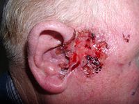
The utility of preoperative and perioperative dermoscopy in standard surgical excision for radical excision of primary basal cell carcinoma remain unexplored. To evaluate the use of preoperative and perioperative dermoscopy for precise mapping of margins during standard surgical excision of primary basal cell carcinoma. In this retrospective, observational study, 17 patients clinically diagnosed with various morphological subtypes of basal cell carcinoma were included. Data about previous history, clinical examination of lesions and regional lymph nodes and preoperative dermoscopy were retrieved. After standard surgical excision had been carried out as per mapping of lateral margins, all the excised surgical specimens were subjected to perioperative dermoscopy and later reconfirmed with histopathology. Seventeen patients with mean age of 60.82 ± 9.99 years and median disease duration of 14 months were analysed. Clinically, basal cell carcinomas were of pigmented superficial subtype [6 (35.3%)], followed by pigmented nodular [5 (29.4%)], nodulo-ulcerative [4 (23.5%)] and micro nodular [2 (11.8%)]. Mean extension of clinical margin after dermoscopy was 0.59 ± 0.52 mm. Mean pre-assessed depth of tumour and mean depth of tumour were 3.46 ± 0.89 mm and 3.49 ± 0.92 mm, respectively. No recurrence was reported. Frequently found pre-operative dermoscopic features were maple leaf like structures [6 (35%)], blue grey dots and globules [6 (35%)] and short fine telangiectasias [6 (35%)]. Commonly observed perioperative dermoscopic features were: (1) irregular band with brown–grey pigmentation of dots, globules, streaks and pseudopodia like extensions [3 (50%)]; (2) irregular band of pseudo granulomatous structureless vascular areas in psoriasiform pattern with diffuse white streaks in pseudopodia like manner [1 (50%)]; (3) irregular band of pseudo granulomatous structureless vascular areas in psoriasiform pattern with streaks of white pseudopodia like structureless areas [1 (50%)]. This was a single-centre study with a small sample size. This study highlights significance of preoperative and perioperative dermoscopy for precise planning and radical excision of primary basal cell carcinoma by standard surgical excision.
Journal Title: Indian Journal of Dermatology, Venereology and Leprology
Year Published: 2023
Link to full text (if available)
Share on Social Media: Sign Up to like & get
recommendations!