Photo from wikipedia
OBJECTIVE Recurrent tonsillitis and obstructive tonsillar hypertrophy are very common in childhood and constitute the two major causes of tonsillectomy in this age group. There is no study in the… Click to show full abstract
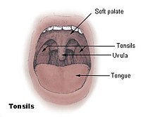
OBJECTIVE Recurrent tonsillitis and obstructive tonsillar hypertrophy are very common in childhood and constitute the two major causes of tonsillectomy in this age group. There is no study in the literature on the immune/histopathological changes in the recurrent and obstructive tonsillar hypertrophy of Weber's glands. In this study, we aimed to histopathologically and immunohistochemically examine the Weber's glands of pediatric patients with recurrent. PATIENTS AND METHODS A total of 63 patients, with 31 patients aged 6-9 who had surgery for recurrent tonsillitis, and 32 patients aged 6-11 years who had surgery for obstructive tonsillar hypertrophy, were included in the study. The removed Weber's glands were included in the obstructive tonsillar hypertrophy or recurrent tonsillitis group according to the patient's clinical diagnosis. All specimens were coded with a numbering method, where only the surgeon knew which patient was in which group. All specimens were evaluated in the same histology center and by the same histologist, unaware of the clinical diagnosis of the patients (blind). RESULTS The comparison of Weber's gland immunohistochemical parameter scores of the groups revealed that the scores of the RT group were significantly higher for all three parameters (VEGF: t=6.777; p<0.001), (EGFR: t=4.386; p<0.001), (IL-6: t=5.072; p<0.001). The comparison of the groups in terms of inflammation, basement membrane thickening, myoepithelial cell and glycoprotein accumulation revealed significantly higher Weber's gland evaluation scores in the RT group for all four parameters. (inflammation: t=7.794; p<0.001), (basement membrane thickening: t=6.582; p<0.001), [myoepithelial cell: t=3.693; p<0.001), (glycoprotein accumulation: t=5.287; p<0.001)]. CONCLUSIONS Histopathological and immunohistochemical examination of Weber's gland in pediatric recurrent tonsillitis and obstructive tonsillar hypertrophy cases revealed inflammatory changes in both disease groups. As expected, inflammatory manifestations were more common in the recurrent tonsillitis group. Besides, inflammatory changes detected in Weber's glands of obstructive tonsillar hypertrophy cases without a history of tonsillitis may contribute to the Weber's gland hypothesis, which attempts to explain the etiology of peritonsillar abscess.
Journal Title: European review for medical and pharmacological sciences
Year Published: 2022
Link to full text (if available)
Share on Social Media: Sign Up to like & get
recommendations!