Photo from wikipedia
A man aged 85 years presented to his treating doctor for his annual skin examination. He had extensive sun damage and multiple previous basal cell carcinomas (BCCs) and squamous cell… Click to show full abstract
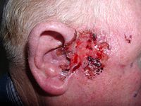
A man aged 85 years presented to his treating doctor for his annual skin examination. He had extensive sun damage and multiple previous basal cell carcinomas (BCCs) and squamous cell carcinomas (SCCs). Examination revealed a lesion on the posterior aspect of his right deltoid (Figure 1). The patient stated that the lesion had been present for decades and that he had not noticed any recent change in size or appearance. The lesion was irregularly shaped, erythematous and slightly sunken, measuring 8 mm in diameter. It had a smooth surface with no scale and was firm to palpation. When squeezed between the thumb and forefinger, the lesion demonstrated a positive ‘delling’ or ‘dimple’ sign, characteristic of a dermatofibroma.1 Close examination revealed milia and erythema between 12 and 3 o’clock.
Journal Title: Australian journal of general practice
Year Published: 2020
Link to full text (if available)
Share on Social Media: Sign Up to like & get
recommendations!