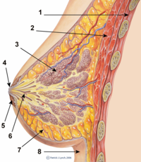
Photo from wikipedia
Intramammary infections in nonlactating mammary glands are common and can occur during periods of rapid mammary epithelial cell (MEC) accumulation, which may ultimately reduce total MEC numbers. Reduced MEC numbers,… Click to show full abstract
Intramammary infections in nonlactating mammary glands are common and can occur during periods of rapid mammary epithelial cell (MEC) accumulation, which may ultimately reduce total MEC numbers. Reduced MEC numbers, resulting from impaired MEC proliferation and increased cellular apoptosis, are expected to reduce future milk yields. The objective of this study was to measure the degree of cellular proliferation and apoptosis in the epithelial and stromal compartment of uninfected and Staphylococcus aureus-infected mammary glands hormonally induced to grow rapidly. Nonpregnant heifers (n = 8) between 11 and 14 mo of age were administered supraphysiological injections of estradiol and progesterone for 14 d. One mammary gland of each heifer was randomly selected and infused with Staph. aureus (CHALL) while another mammary gland was designated as an uninfected control on d 8 of injections. Mammary tissues were collected on the last day of hormonal injections from center and edge parenchymal regions and subject to proliferation assessment via Ki-67 staining and apoptotic assessment via terminal deoxynucleotidyl transferase dUTP nick-end labeling. Differences in cellular proliferation between CHALL and uninfected control quarters were not apparent, but proliferation of MEC was marginally greater in edge parenchyma than in center parenchyma. Coincidently, CHALL quarters experienced a greater percentage of apoptotic MEC and lower percentage of stromal cells undergoing apoptosis than uninfected control quarters. This study also provides the first insight into the mechanisms that allow the mammary fat pad to be replaced by expanding mammary epithelium as edge parenchyma contained a greater percentage of apoptotic stromal cells than center parenchyma. When taken together, these data suggest that Staph. aureus intramammary infection impairs mammary epithelial growth through reductions in MEC number and by preventing its expansion into the mammary fat pad. These factors during periods of rapid mammary growth are expected to impair first lactation milk yield.
Journal Title: Journal of dairy science
Year Published: 2023
Link to full text (if available)
Share on Social Media: Sign Up to like & get
recommendations!