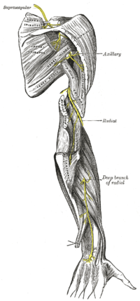
Photo from wikipedia
TO THE EDITOR: We read with great interest the article by Toia et al.4 (Toia F, Gagliardo A, D’Arpa S, et al: Preoperative evaluation of peripheral nerve injuries: What is… Click to show full abstract
TO THE EDITOR: We read with great interest the article by Toia et al.4 (Toia F, Gagliardo A, D’Arpa S, et al: Preoperative evaluation of peripheral nerve injuries: What is the place for ultrasound? J Neurosurg 125:603–614, September 2016). A series of 108 patients with 119 injuries was presented: 69 were affected by nerve entrapment syndromes, 36 by posttraumatic or postsurgical nerve injuries, and only 3 by tumors. We agree with the authors that high-definition ultrasound is a tremendous tool for studying damaged nerves. In fact, it is commonly used in presurgical evaluations, as demonstrated by the numerous clinical articles cited by the authors. The surgical study by Toia et al.4 is noteworthy and prompted some considerations. First, we truthfully think that in entrapment syndromes, ultrasound can be exceptionally useful for planning surgeries, making surgery faster and easier and improving discussions with the patient. But a bifid nerve in a carpal tunnel, for example, does not alter the procedure, and the same can be said for other entrapment syndromes. At the present time, we think that the most important discussion on the role of ultrasound as regards entrapment syndromes is if this tool would, in the future, almost completely replace the more uncomfortable electrophysiological examination: diagnosis of the most common entrapment syndromes could then be based on clinical symptoms and ultrasound findings only. Second, we agree with the authors that the main contribution of ultrasound is its ability to visualize nerve continuity. Ultrasound can distinguish between interrupted fascicles and those in continuity, allowing early differentiation of neurotmetic from axonotmetic injury. For this reason, we consider ultrasound to be mostly useful in posttraumatic and postsurgical nerve injury. Before surgery, ultrasound can even be revolutionary because it allows us to overcome one of the most important problems in the field of peripheral nerve repair—that is, surgical timing. In fact, it is not rare in traumatic peripheral nerve injuries for the surgeon to have difficulty distinguishing between neurotmesis and axonotmesis.1 In these cases, waiting 6 months for an eventual spontaneous functional recovery before performing exploratory surgery is an old rule. In fact, ultrasound actually makes a difference not only in the assessment of posttraumatic nerve injuries, avoiding any delay in treatment, but also in the surgical planning, making surgery faster and easier. In fact, the nerve route is followed with the ultrasound probe until the neuroma and beyond it: taking into account the cross-sectional area and the echoic features of the injured nerve, the gap requiring graft placement can be precisely measured. In this way, tailored skin incisions are drawn over the injury site as well as the donor nerve site, and surgery is more thorough and less time consuming.2 We also want to emphasize the role of ultrasound after reconstructive surgery. In fact, during follow-up, the graft shape can be easily checked, and the eventual scar tissue as well as its misplacement can be detected early.2 The third consideration is related to the role of ultrasound in peripheral nerve tumors, most of which are schwannomas. In this field, we think that the most important diagnostic “tool” in the majority of cases is the clinical examination together with the survey for tumefaction and a Tinel sign: ultrasound and/or other radiological examinations such as MRI and/or CT support the surgeon in confirming the suspected diagnosis. Then, standard microsurgery allows selective and safe tumor removal, after recognizing and dissecting most of the nerve fascicles not involved by the tumor.3 In conclusion, we think that ultrasound can be really useful for improving surgical planning, decision making, and clinical follow-up and that in the near future, with the finest techniques, ultrasound could replace the use of neurophysiological studies in peripheral nerve injuries.
Journal Title: Journal of neurosurgery
Year Published: 2017
Link to full text (if available)
Share on Social Media: Sign Up to like & get
recommendations!