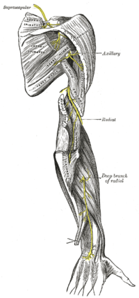
Photo from wikipedia
OBJECTIVE The authors describe the anatomy of the motor branches of the pronator teres (PT) as it relates to transferring the nerve of the extensor carpi radialis brevis (ECRB) to… Click to show full abstract
OBJECTIVE The authors describe the anatomy of the motor branches of the pronator teres (PT) as it relates to transferring the nerve of the extensor carpi radialis brevis (ECRB) to restore wrist extension in patients with radial nerve paralysis. They describe their anatomical cadaveric findings and report the results of their nerve transfer technique in several patients followed for at least 24 months postoperatively. METHODS The authors dissected both upper limbs of 16 fresh cadavers. In 6 patients undergoing nerve surgery on the elbow, they dissected the branches of the median nerve and confirmed their identity by electrical stimulation. Of these 6 patients, 5 had had a radial nerve injury lasting 7-12 months, underwent transfer of the distal PT motor branch to the ECRB, and were followed for at least 24 months. RESULTS The PT was innervated by two branches: a proximal branch, arising at a distance between 0 and 40 mm distal to the medial epicondyle, responsible for PT superficial head innervation, and a distal motor branch, emerging from the anterior side of the median nerve at a distance between 25 and 60 mm distal to the medial epicondyle. The distal motor branch of the PT traveled approximately 30 mm along the anterior side of the median nerve; just before the median nerve passed between the PT heads, it bifurcated to innervate the deep head and distal part of the superficial head of the PT. In 30% of the cadaver limbs, the proximal and distal PT branches converged into a single trunk distal to the medial epicondyle, while they converged into a single branch proximal to it in 70% of the limbs. The proximal and distal motor branches of the PT and the nerve to the ECRB had an average of 646, 599, and 457 myelinated fibers, respectively.All patients recovered full range of wrist flexion-extension, grade M4 strength on the British Medical Research Council scale. Grasp strength recovery achieved almost 50% of the strength of the contralateral side. All patients could maintain their wrist in extension while performing grasp measurements. CONCLUSIONS The distal PT motor branch is suitable for reinnervation of the ECRB in radial nerve paralysis, for as long as 7-12 months postinjury.
Journal Title: Journal of neurosurgery
Year Published: 2020
Link to full text (if available)
Share on Social Media: Sign Up to like & get
recommendations!