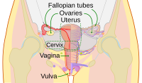
Photo from wikipedia
Vaginal mucosal surfaces naturally offer some protection against sexually transmitted infections (STIs) including Human Immunodeficiency Virus-1, however topical preventative medications or vaccine designed to boost local immune responses can further… Click to show full abstract
Vaginal mucosal surfaces naturally offer some protection against sexually transmitted infections (STIs) including Human Immunodeficiency Virus-1, however topical preventative medications or vaccine designed to boost local immune responses can further enhance this protection. We previously developed a novel mucosal vaccine strategy using viral vectors integrated into mouse dermal epithelium to induce virus-specific humoral and cellular immune responses at the site of exposure. Since vaccine integration occurs at the site of cell replication (basal layer 100-400 micrometers below the surface), temporal epithelial thinning during vaccine application, confirmed with high resolution imaging, is desirable. In this study, strategies for vaginal mucosal thinning were evaluated noninvasively using optical coherence tomography (OCT) to map reproductive tract epithelial thickness (ET) in macaques to optimize basal layer access in preparation for future effective intravaginal mucosal vaccination studies. Twelve adolescent female rhesus macaques (5-7kg) were randomly assigned to interventions to induce vaginal mucosal thinning, including cytobrush mechanical abrasion, the chemical surfactant spermicide nonoxynol-9 (N9), the hormonal contraceptive depomedroxyprogesterone acetate (DMPA), or no intervention. Macaques were evaluated at baseline and after interventions using colposcopy, vaginal biopsies, and OCT imaging, which allowed for real-time in vivo visualization and measurement of ET of the mid-vagina, fornices, and cervix. P value ≤0.05 was considered significant. Colposcopy findings included pink, rugated tissue with variable degrees of white-tipped, thickened epithelium. Baseline ET of the fornices was thinner than the cervix and vagina (p<0.05), and mensing macaques had thinner ET at all sites (p<0.001). ET was decreased 1 month after DMPA (p<0.05) in all sites, immediately after mechanical abrasion (p<0.05) in the fornix and cervix, and after two doses of 4% N9 (1.25ml) applied over 14 hrs in the fornix only (p<0.001). Histological assessment of biopsied samples confirmed OCT findings. In summary, OCT imaging allowed for real time assessment of macaque vaginal ET. While varying degrees of thinning were observed after the interventions, limitations with each were noted. ET decreased naturally during menses, which may provide an ideal opportunity for accessing the targeted vaginal mucosal basal layers to achieve the optimum epithelial thickness for intravaginal mucosal vaccination.
Journal Title: Frontiers in Immunology
Year Published: 2021
Link to full text (if available)
Share on Social Media: Sign Up to like & get
recommendations!