Photo from wikipedia
Purpose To investigate changes in retinal circulation and the choroid in patients with acute myeloid leukemia (AML) in the acute and remission stages, to analyze the correlation between retinal circulation… Click to show full abstract
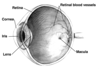
Purpose To investigate changes in retinal circulation and the choroid in patients with acute myeloid leukemia (AML) in the acute and remission stages, to analyze the correlation between retinal circulation and laboratory parameters, and to assess risk factors associated with leukemic retinopathy. Methods Forty-eight patients (93 eyes) with AML were enrolled and divided into two groups according to fundus examination findings: the retinopathy and no retinopathy groups. Patients underwent eye measurements before treatment and after remission. Macular vessel density (VD), perfusion density (PD), foveal avascular zone (FAZ), and choroidal thickness (ChT) were measured using optical coherence tomography angiography. Patients with healthy eyes were recruited as control participants. Results Patients with leukemic retinopathy had higher measurements of white blood cells (WBCs), circulating blasts, fibrin degradation products, and cross-linked fibrin degradation products (D-dimer) and a lower hemoglobin (HB) count (p < 0.05). In the acute phase of the disease, the VD and PD were lower and the ChT was thicker in patients with AML than in controls (p < 0.05), irrespective of the presence of leukemic retinopathy; however, the patients were partially recovered in the remission stage. The VD was lower in patients with higher WBC (r = −0.217, p = 0.036), D-dimer (r = −0.279, p = 0.001), fasting blood glucose (FBG) (r = −0.298, p = 0.004) and triglyceride (r = −0.336, p = 0.001) levels. The FAZ area was negatively correlated with HB (r = −0.258, p = 0.012). Conclusion Patients with AML appear to have subclinical retinal perfusion loss and choroidal thickening in the acute phase of the disease, but this is reversible. Injury to bone marrow function may cause a decrease in retinal perfusion. Leukemic retinopathy is associated with abnormal hematologic parameters and coagulopathy.
Journal Title: Frontiers in Medicine
Year Published: 2023
Link to full text (if available)
Share on Social Media: Sign Up to like & get
recommendations!