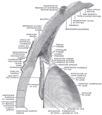
Photo from wikipedia
Objective This study aimed to investigate the microscopic structure and characteristics of nevi on the conjunctiva of the lacrimal caruncle by in vivo confocal microscopy. Methods In total, four patients… Click to show full abstract
Objective This study aimed to investigate the microscopic structure and characteristics of nevi on the conjunctiva of the lacrimal caruncle by in vivo confocal microscopy. Methods In total, four patients with nevi growing on the lacrimal caruncle conjunctiva were recruited. The morphological characteristics of the nevi were evaluated by in vivo confocal microscopy before excision surgery; the results were compared with histopathological analyses of the surgical specimens. Results The nevi of the four patients were all located at the conjunctiva of the lacrimal caruncle, with a slightly nodular surface, mixed black and brown color, and clear boundary. The nevi were round and highly protruded on the surface of the lacrimal caruncle, with an average diameter of 4.5 ± 1.29 mm. Under in vivo confocal microscopy, the pigmented nevus cells on the conjunctiva of the lacrimal caruncle were observed to be clustered in nests with irregular boundaries. The cells were round or irregular, with clear cell boundaries, hyper-reflective at the periphery, with low reflectivity in the center. Vascular crawling was observed in some regions. Histopathological analysis showed that nevus cells were roughly equal in size and distributed in a nodular pattern. Melanin granules were observed in the cytoplasm. No atypia or mitotic figures of the cells were found. Conclusion This study revealed that the microstructure of nevi growing on the conjunctiva of the lacrimal caruncle can be identified by in vivo confocal microscopy.
Journal Title: Frontiers in Medicine
Year Published: 2023
Link to full text (if available)
Share on Social Media: Sign Up to like & get
recommendations!