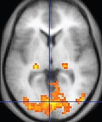
Photo from wikipedia
Resting-state functional magnetic images (rs-fMRIs) can be used to map and delineate the brain activity occurring while the patient is in a task-free state. These resting-state activity networks can be… Click to show full abstract
Resting-state functional magnetic images (rs-fMRIs) can be used to map and delineate the brain activity occurring while the patient is in a task-free state. These resting-state activity networks can be informative when diagnosing various neurodevelopmental diseases, but only if the images are high quality. The quality of an rs-fMRI rapidly degrades when the patient moves during the scan. Herein, we describe how patient motion impacts an rs-fMRI on multiple levels. We begin with how the electromagnetic field and pulses of an MR scanner interact with a patient’s physiology, how movement affects the net signal acquired by the scanner, and how motion can be quantified from rs-fMRI. We then present methods for preventing motion through educational and behavioral interventions appropriate for different age groups, techniques for prospectively monitoring and correcting motion during the acquisition process, and pipelines for mitigating the effects of motion in existing scans.
Journal Title: Diagnostics
Year Published: 2022
Link to full text (if available)
Share on Social Media: Sign Up to like & get
recommendations!