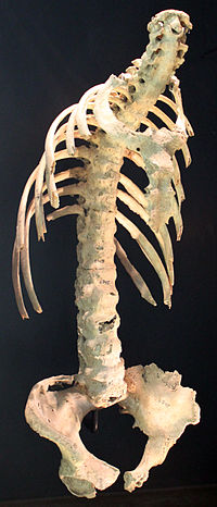
Photo from wikipedia
Diffuse idiopathic skeletal hyperostosis (DISH) is a systemic condition characterized by new bone formation and enthesopathies of the axial and peripheral skeleton. The pathogenesis of DISH is not well understood,… Click to show full abstract
Diffuse idiopathic skeletal hyperostosis (DISH) is a systemic condition characterized by new bone formation and enthesopathies of the axial and peripheral skeleton. The pathogenesis of DISH is not well understood, and it is currently considered a non-inflammatory condition with an underlying metabolic derangement. Currently, DISH diagnosis relies on the Resnick and Niwayama criteria, which encompass end-stage disease with an already ankylotic spine. Imaging characterization of the axial and peripheral skeleton in DISH subjects may potentially help identify earlier diagnostic criteria and provide further data for deciphering the general pathogenesis of DISH and new bone formation. In the current review, we aim to summarize and characterize axial and peripheral imaging findings of the skeleton related to DISH, along with their clinical and pathogenetic relevance.
Journal Title: Diagnostics
Year Published: 2023
Link to full text (if available)
Share on Social Media: Sign Up to like & get
recommendations!