Photo from wikipedia
Tumor cells evolve in a complex and heterogeneous environment composed of different cell types and an extracellular matrix. Current 2D culture methods are very limited in their ability to mimic… Click to show full abstract
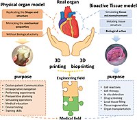
Tumor cells evolve in a complex and heterogeneous environment composed of different cell types and an extracellular matrix. Current 2D culture methods are very limited in their ability to mimic the cancer cell environment. In recent years, various 3D models of cancer cells have been developed, notably in the form of spheroids/organoids, using scaffold or cancer-on-chip devices. However, these models have the disadvantage of not being able to precisely control the organization of multiple cell types in complex architecture and are sometimes not very reproducible in their production, and this is especially true for spheroids. Three-dimensional bioprinting can produce complex, multi-cellular, and reproducible constructs in which the matrix composition and rigidity can be adapted locally or globally to the tumor model studied. For these reasons, 3D bioprinting seems to be the technique of choice to mimic the tumor microenvironment in vivo as closely as possible. In this review, we discuss different 3D-bioprinting technologies, including bioinks and crosslinkers that can be used for in vitro cancer models and the techniques used to study cells grown in hydrogels; finally, we provide some applications of bioprinted cancer models.
Journal Title: International Journal of Molecular Sciences
Year Published: 2022
Link to full text (if available)
Share on Social Media: Sign Up to like & get
recommendations!