Photo from wikipedia
Fascin, a major actin cross-linking protein, is expressed in most vertebrate epithelial tissues. It organizes actin filaments into well-ordered bundles that are responsible for the extension of dynamic membrane protrusions,… Click to show full abstract
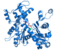
Fascin, a major actin cross-linking protein, is expressed in most vertebrate epithelial tissues. It organizes actin filaments into well-ordered bundles that are responsible for the extension of dynamic membrane protrusions, including microspikes, filopodia, and invadopodia from cell surfaces, which are involved in cell migration and invasion as critical components of cancer metastasis. However, it is not well-understood how fascin-1 induces actin binding/bundling and where fascin-1 localizes along the actin filaments, thus facilitating actin bundle formation. In the present study, we attempted to clarify these problems by using biochemical and electron microscopic analyses using various fascin-1 constructs. Three dimensional structures of actin/fascin-1 complex were obtained by electron microscopy (EM) with iterative helical real-space reconstruction (IHRSR) and tomography. We revealed that the N-terminal region containing the Actin-Binding Site 2 (ABS2) of fascin-1 is responsible for actin bundling and the C-terminal region is important for the dimerization of fascin-1. We also found that the dimerization of fascin-1 through intermolecular interactions of the C-terminal region is essential for actin bundling. Since fascin is an important factor in cancer development, it is expected that the findings of present study will provide useful information for development of therapeutic strategies for cancer.
Journal Title: Life
Year Published: 2022
Link to full text (if available)
Share on Social Media: Sign Up to like & get
recommendations!