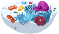
Photo from wikipedia
The metabolic basis of Parkinson’s disease pathology is poorly understood. However, the involvement of mitochondrial and endoplasmic reticulum stress in dopamine neurons in disease aetiology is well established. We looked… Click to show full abstract
The metabolic basis of Parkinson’s disease pathology is poorly understood. However, the involvement of mitochondrial and endoplasmic reticulum stress in dopamine neurons in disease aetiology is well established. We looked at the effect of rotenone- and tunicamycin-induced mitochondrial and ER stress on the metabolism of wild type and microtubule-associated protein tau mutant dopamine neurons. Dopamine neurons derived from human isolated iPSCs were subjected to mitochondrial and ER stress using RT and TM, respectively. Comprehensive metabolite profiles were generated using a split phase extraction analysed by reversed phase lipidomics whilst the aqueous phase was measured using HILIC metabolomics. Mitochondrial and ER stress were both shown to cause significant dysregulation of metabolism with RT-induced stress producing a larger shift in the metabolic profile of both wild type and MAPT neurons. Detailed analysis showed that accumulation of triglycerides was a significant driver of metabolic dysregulation in response to both stresses in both genotypes. Whilst the consequence is similar, the mechanisms by which triglyceride accumulation occurs in dopamine neurons in response to mitochondrial and ER stress are very different. Thus, improving our understanding of how these mechanisms drive the observed triglyceride accumulation can potentially open up new therapeutic avenues.
Journal Title: Metabolites
Year Published: 2023
Link to full text (if available)
Share on Social Media: Sign Up to like & get
recommendations!