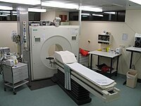
Photo from wikipedia
Dysregulation of microtubules is commonly associated with several psychiatric and neurological disorders, including addiction and Alzheimer’s disease. Imaging of microtubules in vivo using positron emission tomography (PET) could provide valuable… Click to show full abstract
Dysregulation of microtubules is commonly associated with several psychiatric and neurological disorders, including addiction and Alzheimer’s disease. Imaging of microtubules in vivo using positron emission tomography (PET) could provide valuable information on their role in the development of disease pathogenesis and aid in improving therapeutic regimens. We developed [11C]MPC-6827, the first brain-penetrating PET radiotracer to image microtubules in vivo in the mouse brain. The aim of the present study was to assess the reproducibility of [11C]MPC-6827 PET imaging in non-human primate brains. Two dynamic 0–120 min PET/CT imaging scans were performed in each of four healthy male cynomolgus monkeys approximately one week apart. Time activity curves (TACs) and standard uptake values (SUVs) were determined for whole brains and specific regions of the brains and compared between the “test” and “retest” data. [11C]MPC-6827 showed excellent brain uptake with good pharmacokinetics in non-human primate brains, with significant correlation between the test and retest scan data (r = 0.77, p = 0.023). These initial evaluations demonstrate the high translational potential of [11C]MPC-6827 to image microtubules in the brain in vivo in monkey models of neurological and psychiatric diseases.
Journal Title: Molecules
Year Published: 2020
Link to full text (if available)
Share on Social Media: Sign Up to like & get
recommendations!