Photo from wikipedia
Focusing on the transmembrane domains (TMDs) of viral fusion and channel-forming proteins (VCPs), experimentally available and newly generated peptides in an ideal conformation of the S and E proteins of… Click to show full abstract
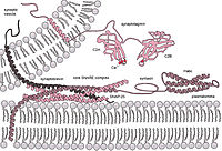
Focusing on the transmembrane domains (TMDs) of viral fusion and channel-forming proteins (VCPs), experimentally available and newly generated peptides in an ideal conformation of the S and E proteins of severe acute respiratory syndrome coronavirus type 2 (SARS-CoV-2) and SARS-CoV, gp41 and Vpu, both of human immunodeficiency virus type 1 (HIV-1), haemagglutinin and M2 of influenza A, as well as gB of herpes simplex virus (HSV), are embedded in a fully hydrated lipid bilayer and used in multi-nanosecond molecular dynamics simulations. It is aimed to identify differences in the dynamics of the individual TMDs of the two types of viral membrane proteins. The assumption is made that the dynamics of the individual TMDs are decoupled from their extra-membrane domains, and that the mechanics of the TMDs are distinct from each other due to the different mechanism of function of the two types of proteins. The diffusivity coefficient (DC) of the translational and rotational diffusion is decreased in the oligomeric state of the TMDs compared to those values when calculated from simulations in their monomeric state. When comparing the calculations for two different lengths of the TMD, a longer full peptide and a shorter purely TMD stretch, (i) the difference of the calculated DCs begins to level out when the difference exceeds approximately 15 amino acids per peptide chain, and (ii) the channel protein rotational DC is the most affected diffusion parameter. The rotational dynamics of the individual amino acids within the middle section of the TMDs of the fusion peptides remain high upon oligomerization, but decrease for the channel peptides, with an increasing number of monomers forming the oligomeric state, suggesting an entropic penalty on oligomerization for the latter.
Journal Title: Viruses
Year Published: 2022
Link to full text (if available)
Share on Social Media: Sign Up to like & get
recommendations!