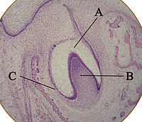
Photo from wikipedia
FUN14 domain‑containing 1 (FUNDC1) is a receptor that has been previously reported to activate hypoxia‑induced mitophagy. However, the potential role of FUNDC1 in the pathophysiology of dental pulp diseases remains… Click to show full abstract
FUN14 domain‑containing 1 (FUNDC1) is a receptor that has been previously reported to activate hypoxia‑induced mitophagy. However, the potential role of FUNDC1 in the pathophysiology of dental pulp diseases remains unknown. Therefore, present study first collected tissue specimens from patients with pulpitis and from healthy individuals. The results of reverse transcription‑quantitative PCR and immunohistochemical staining revealed markedly increased FUNDC1 and hypoxia‑inducible factor‑1α expression in pulpitis tissue specimens compared with those from healthy individuals. To provide a theoretical basis for the study of the occurrence, development and reparative mechanisms in the dental pulp after tissue injury, the present study then investigated the role of hypoxia‑induced mitophagy in the regulation of proliferation, migration and odontoblastic differentiation in human dental pulp cells (HDPCs), in addition, to the possible involvement of FUNDC1. The surface markers and multipotent differentiation capabilities of HDPCs were performed by flow cytometry (surface markers), alizarin red (osteogenic capabilities), alcian blue (chondrogenic capabilities) and oil red O (adipogenic capabilities). Following culture under hypoxia conditions (1% O2) for varying time periods, the proliferation, migration and odontoblastic differentiation of HDPCs were measured using Cell Counting Kit‑8, wound healing and Transwell migration assays, alkaline phosphatase staining and activity tests and western blotting (runt‑related transcription factor 2, collagen I, osterix and osteopontin), respectively. Immunofluorescence and western blotting were performed to measure the expression levels of hypoxia‑inducible factor‑1α, pro‑fission dynamin‑related protein 1, mitochondria‑related proteins translocase of inner mitochondrial membrane 23 and translocase of outer mitochondrial membrane 20, in addition to those of autophagy markers (p62, LC3II, Beclin‑1 and autophagy‑related 5). Transmission electron microscopy was also used to image the autophagosomes and mitochondrial morphology. In addition, to study the functional role of FUNDC1, its expression was silenced by liposome‑mediated transfection with small interfering RNA into HDPCs. Compared with those in HDPCs cultured under normoxic conditions (21% O2), the ability of autophagy in HDPCs cultured under hypoxic conditions for 18 h was markedly increased, whilst the proliferation, migration and odontoblastic differentiation were also enhanced. Increased numbers of autophagosomes could also be observed in the hypoxic group. However, FUNDC1 knockdown in HDPCs reversed the aforementioned effects. Overall, data from the present study suggest that hypoxia can promote the proliferation, migration and odontoblastic differentiation of HDPCs, where the underlying mechanism may be associated with the activation of mitophagy downstream of FUNDC1.
Journal Title: International journal of molecular medicine
Year Published: 2022
Link to full text (if available)
Share on Social Media: Sign Up to like & get
recommendations!